
پرونده:Gray760.png
Gray760.png (۵۰۰ × ۳۲۳ پیکسل، اندازهٔ پرونده: ۲۳ کیلوبایت، نوع MIME پرونده: image/png)
این پرونده در ویکیانبار موجود است. محتویات صفحهٔ توصیف آن در زیر نمایش داده میشود. |
خلاصه
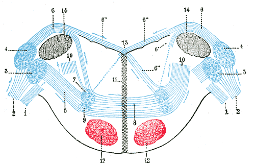
Terminal nuclei of the cochlear nerve, with their upper connections. (Schematic.) The vestibular nerve with its terminal nuclei and their efferent fibers have been suppressed. On the other hand, in order not to obscure the trapezoid body, the efferent fibers of the terminal nuclei on the right side have been resected in a considerable portion of their extent. The trapezoid body, therefore, shows only one-half of its fibers, viz., those which come from the left.
1. Vestibular nerve, divided at its entrance into the medulla oblongata.
2. Cochlear nerve.
3. Accessory nucleus of acoustic nerve.
4. Tuberculum acusticum.
5. Efferent fibers of accessory nucleus.
6. Efferent fibers of tuberculum acusticum, forming the striae medullares, with 6’, their direct bundle going to the superior olivary nucleus of the same side; 6’’, their decussating bundles going to the superior olivary nucleus of the opposite side.
7. Superior olivary nucleus.
8. Trapezoid body.
9. Trapezoid nucleus.
10. Central acoustic tract (lateral lemniscus).
11. Raphé.
12. Cerebrospinal fasciculus.
13. Fourth ventricle.
14. Inferior peduncle.
| توضیح | Terminal nuclei of the cochlear nerve, with their upper connections. (Schematic.) The vestibular nerve with its terminal nuclei and their efferent fibers have been suppressed. On the other hand, in order not to obscure the trapezoid body, the efferent fibers of the terminal nuclei on the right side have been resected in a considerable portion of their extent. The trapezoid body, therefore, shows only one-half of its fibers, viz., those which come from the left. 1. Vestibular nerve, divided at its entrance into the medulla oblongata. 2. Cochlear nerve. 3. Accessory nucleus of acoustic nerve. 4. Tuberculum acusticum. 5. Efferent fibers of accessory nucleus. 6. Efferent fibers of tuberculum acusticum, forming the striae medullares, with 6’, their direct bundle going to the superior olivary nucleus of the same side; 6’’, their decussating bundles going to the superior olivary nucleus of the opposite side. 7. Superior olivary nucleus. 8. Trapezoid body. 9. Trapezoid nucleus. 10. Central acoustic tract (lateral lemniscus). 11. Raphé. 12. Cerebrospinal fasciculus. 13. Fourth ventricle. 14. Inferior peduncle. (Testut.) | ||||||||||||||||||||
| Plate | 760 | ||||||||||||||||||||
| تاریخ | پیش از ۱۸۵۸ | ||||||||||||||||||||
| منبع |
|
||||||||||||||||||||
| پدیدآور |
|
||||||||||||||||||||
.jpg)
کتاب
| هنری گری: Gray's Anatomy (20th edition)
|
|||||||||||||||||||||||
|---|---|---|---|---|---|---|---|---|---|---|---|---|---|---|---|---|---|---|---|---|---|---|---|
| پدیدآور |
|
-_Title_page.png) | |||||||||||||||||||||
| ویرایشگر |
Revised by Warren H. Lewis |
||||||||||||||||||||||
| تصویرگر |
|
||||||||||||||||||||||
| عنوان | |||||||||||||||||||||||
| نسخه |
20 |
||||||||||||||||||||||
| ناشر | |||||||||||||||||||||||
| نوع شیء |
ویراست |
||||||||||||||||||||||
| بازنگری صفحه | list of all the plates | ||||||||||||||||||||||
| زبان |
زبان انگلیسی |
||||||||||||||||||||||
| تاریخ انتشار |
۱۹۱۸ |
||||||||||||||||||||||
| محل انتشار |
فیلادلفیا / نیویورک |
||||||||||||||||||||||
| منبع | Bartleby | ||||||||||||||||||||||

اجازهنامه
This image is in the public domain because it is a mere mechanical scan or photocopy of a public domain original, or – from the available evidence – is so similar to such a scan or photocopy that no copyright protection can be expected to arise. The original itself is in the public domain for the following reason:
This tag is designed for use where there may be a need to assert that any enhancements (eg brightness, contrast, colour-matching, sharpening) are in themselves insufficiently creative to generate a new copyright. It can be used where it is unknown whether any enhancements have been made, as well as when the enhancements are clear but insufficient. For known raw unenhanced scans you can use an appropriate {{PD-old}} tag instead. For usage, see Commons:When to use the PD-scan tag.  | ||||
تاریخچهٔ پرونده
روی تاریخ/زمانها کلیک کنید تا نسخهٔ مربوط به آن هنگام را ببینید.
| تاریخ/زمان | بندانگشتی | ابعاد | کاربر | توضیح | |
|---|---|---|---|---|---|
| کنونی | ۲۳ ژانویهٔ ۲۰۰۷، ساعت ۲۰:۵۲ | | ۵۰۰ در ۳۲۳ (۲۳ کیلوبایت) | Pngbot | optimized with optipng |
| ۲۹ ژانویهٔ ۲۰۰۶، ساعت ۰۵:۳۶ |  | ۵۰۰ در ۳۲۳ (۳۹ کیلوبایت) | Arcadian | {{Gray's Anatomy plate}} |
کاربرد پرونده
صفحههای زیر از این تصویر استفاده میکنند:
کاربرد سراسری پرونده
ویکیهای دیگر زیر از این پرونده استفاده میکنند:
- کاربرد در ar.wikipedia.org
- کاربرد در bg.wikipedia.org
- کاربرد در de.wikipedia.org
- کاربرد در de.wikibooks.org
- کاربرد در en.wikipedia.org
- کاربرد در es.wikipedia.org
- کاربرد در ja.wikipedia.org
- کاربرد در kk.wikipedia.org
- کاربرد در nl.wikipedia.org
- کاربرد در pl.wikipedia.org
- کاربرد در zh.wikipedia.org